Anatomical Name Of Lower Back Muscles / Low Back Pain Wikipedia. Out of these, the cookies that are categorized as necessary are stored on your browser as they are essential for the working of basic functionalities of the website. It also contributes to the ipsilateral lateral flexion of the neck. 1 your spine in this region has a natural inward curve. This website uses cookies to improve your experience while you navigate through the website. Last time we learned the anatomical details of the lower back muscles.
They help to bend the back to one side or the other. The back muscles can be three types. These muscles include the large paired muscles in the lower back, called erector spinae, which help hold up the spine, and gluteal muscles. Each lumbar spinal level is numbered from top to bottom—l1 through l5, or l6. Name the 4 muscles of the quadriceps femoris group.
Thoracic Spine from cloud2.spineuniverse.com A skull consists of the frontal, temporal, parietal and occipital bones. The back consists of the spine, spinal cord, muscles, ligaments, and nerves. The teres major muscle originates on the outer (lateral) edge of the scapula and attaches to the humerus. The teres majo r muscles work with the rotator cuff muscles to stabilize. The muscles of the back that work together to support the spine, help keep the body upright and allow twist and bend in many directions. Balance the weight of your head on top of your spine The flexor muscles are attached to the front of the spine and enable flexing, bending forward, lifting, and arching the lower back. Each lumbar spinal level is numbered from top to bottom—l1 through l5, or l6.
The muscles of the back are a group of strong, paired muscles that lie on the posterior aspect of the trunk they provide movements of the spine, stability to the trunk, as well as the coordination between the movements of the limbs and the back muscles are divided into two large groups:
The vertebral column of the lower back includes the five lumbar vertebrae, the sacrum, and the coccyx. Out of these, the cookies that are categorized as necessary are stored on your browser as they are essential for the working of basic functionalities of the website. The muscles on the back of the trunk help lower the arms and move the body forward and sideways. There are three parts to the trapezius. The back muscles can be three types. Lumbar spine lower back and superficial muscles the muscles of the lower back help stabilize, rotate, flex, and extend the spinal column, which is a bony tower of 24 vertebrae that gives the body. Chart of major posterior muscles. The extrinsic (superficial) back muscles, which lie most superficially on the back. Three types of back muscles that help the spine function are extensors, flexors and obliques. Sometimes the name of the muscle includes it's function—such as extensor, flexor, adductor, abductor. These structures work together to support the body, enable a range of movements, and send messages from the brain to. They also protect the spinal column. The lower part of the trapezius ascends and depresses the scapula, while the transverse or middle region of the trapezius is what retracts the.
Muscles of the lower back and buttocks diagram, human muscles, muscles of the lower back and buttocks diagram. The superficial group, also known as the appendicular group, is primarily associated with movement of the appendicular skeleton. The quadratus lumborum muscles (orange, in the image above) are found in the lower back (also called the lumbar area). The lower leg is a major anatomical part of the skeletal system. Rectus femoris, vastus lateralis, vastus medialis, vastus intermedius.
Low Back Pain A Guide For Coaches And Athletes On Anatomy Types And Treatment Breaking Muscle from upload.wikimedia.org Sometimes the name of the muscle includes it's function—such as extensor, flexor, adductor, abductor. It is innervated by anterior rami of spinal nerves, reflecting its embryological origin outside the back. The upper back is a complex area containing a number of muscles that perform various actions on the scapulae (shoulder blades) and humerus. Muscles of lower back diagram. It lies between the knee and the ankle, while the upper leg lies between. The muscles that move the upper legs (thigh) there are many muscles that move the large bone of the thigh. Together with the upper leg, it forms the lower extremity. Rectus femoris, vastus lateralis, vastus medialis, vastus intermedius.
Sometimes the name of the muscle includes it's function—such as extensor, flexor, adductor, abductor.
It lies between the knee and the ankle, while the upper leg lies between. The muscles that move the upper legs (thigh) there are many muscles that move the large bone of the thigh. The teres major muscle originates on the outer (lateral) edge of the scapula and attaches to the humerus. Anatomical name of lower back muscles | the muscular system consists of the skeletal muscles and their associated structures. Balance the weight of your head on top of your spine It is innervated by anterior rami of spinal nerves, reflecting its embryological origin outside the back. This website uses cookies to improve your experience while you navigate through the website. Related posts of muscle names of lower back muscle anatomy pictures. The lordotic curve your lower back (lumbar spine) is the anatomic region between your lowest rib and the upper part of the buttock. Muscles of lower back diagram. The upper back is a complex area containing a number of muscles that perform various actions on the scapulae (shoulder blades) and humerus. Human musculature bodybuilding infographic muscular system vector human anatomy back muscle anatomy bicep male muscular anatomy human body anatomy female female anatomy muscle hamstrings muscle. Anatomical name of lower back muscles :
The superficial group, also known as the appendicular group, is primarily associated with movement of the appendicular skeleton. The lower part of the trapezius ascends and depresses the scapula, while the transverse or middle region of the trapezius is what retracts the. The muscle then courses up to your shoulder and attaches to your upper arm bone. The teres majo r muscles work with the rotator cuff muscles to stabilize. Each lumbar spinal level is numbered from top to bottom—l1 through l5, or l6.
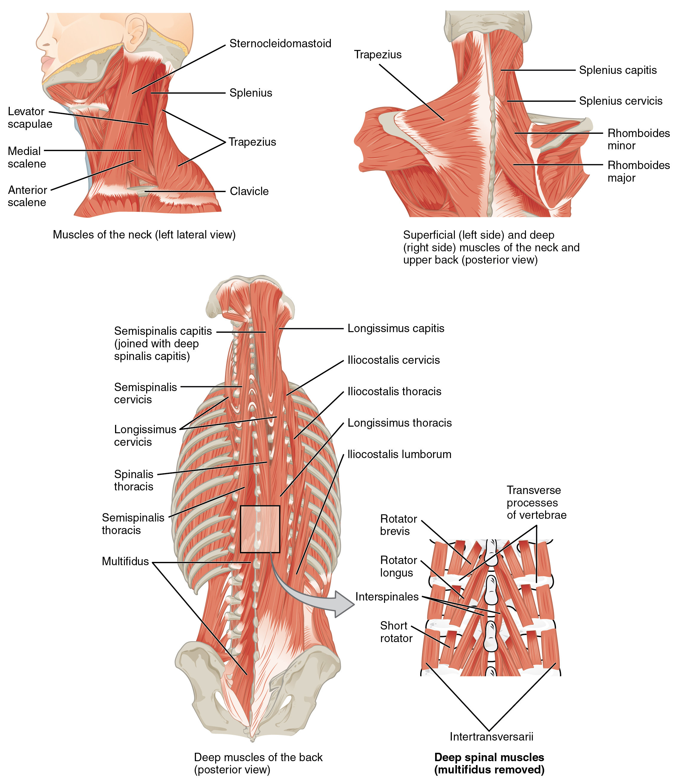
Lumbar Spine Anatomy from www.spineuniverse.com It is composed of trapezius, latissimus dorsi, rhomboid major, rhomboid minor and levator scapulae. Anatomical name of lower back muscles : Sometimes the name of the muscle includes it's function—such as extensor, flexor, adductor, abductor. Three types of back muscles that help the spine function are extensors, flexors and obliques. The quick answer to this question is the muscles of the lower back are the multifidus, longissimus, spinalis, and quadratus lumborum. Muscle anatomy pictures 12 photos of the muscle anatomy pictures female muscle anatomy pictures, human muscle anatomy images download, leg muscle anatomy pictures, pictures of muscle anatomy, quizlet muscle anatomy pictures, human muscles, female muscle anatomy pictures, human muscle anatomy images download, leg muscle. Intermediate back muscles and c. They originate from the thoracolumbar fascia, the spinous process of thoracic six through 12, the iliac crest, and your lower three ribs.
An extremely strong tendon attached to the heel.
Human musculature bodybuilding infographic muscular system vector human anatomy back muscle anatomy bicep male muscular anatomy human body anatomy female female anatomy muscle hamstrings muscle. The back muscles represented on an anatomical chart and on a schematic view of the origin and insertion of the proper muscles of the back (iliocostal muscle of. The muscles that move the upper legs (thigh) there are many muscles that move the large bone of the thigh. The lower leg is a major anatomical part of the skeletal system. Your lats are a major back muscle and mover of your shoulder joint. Out of these, the cookies that are categorized as necessary are stored on your browser as they are essential for the working of basic functionalities of the website. Together with the upper leg, it forms the lower extremity. Rectus femoris, vastus lateralis, vastus medialis, vastus intermedius. Sometimes the name of the muscle includes it's function—such as extensor, flexor, adductor, abductor. These structures work together to support the body, enable a range of movements, and send messages from the brain to. It also contributes to the ipsilateral lateral flexion of the neck. This website uses cookies to improve your experience while you navigate through the website. The teres major muscle originates on the outer (lateral) edge of the scapula and attaches to the humerus.